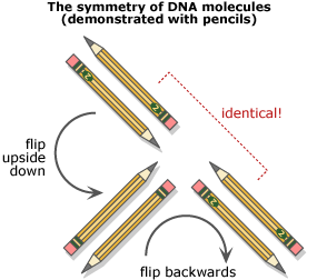
 While Watson and Crick went back to their model building, Franklin continued to work on DNA by making X-ray diffraction images and analyzing these results. She and Gosling focused on DNA A, producing many clear images and uncovering more clues to its structure: the size of the repeating units that made up the molecule and the symmetry of these units. DNA crystals, it turned out, look the same when they are turned upside down and backwards.

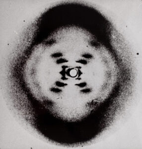
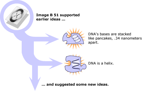
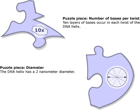
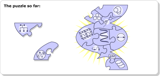
Each image took many hours of X-ray exposure to develop — sometimes up to 100 hours — so Franklin and Gosling occasionally exposed them overnight. On the morning of May 2nd, 1952, they returned to the lab to discover that the DNA had hydrated during the night and the image they had taken was actually of DNA B. It was unusually sharp — and illuminating. It showed an obvious x shape, a pattern that previous work associated with helical structures. The image also confirmed the idea that DNA’s bases were stacked pancake-style, .34 nanometers apart, and suggested that 10 of these layers occurred in every twist of the helix. It even delineated the width of the diameter of the helix: 2 nanometers. Since it was the 51st image taken, they called it image B 51. They set it aside and decided to come back to it once they’d solved the structure of DNA A.
Franklin and Gosling’s production of image B 51 was an accident that would profoundly influence the course of the race for DNA’s structure. To learn more about the role of serendipity in science, visit The story of serendipity.
